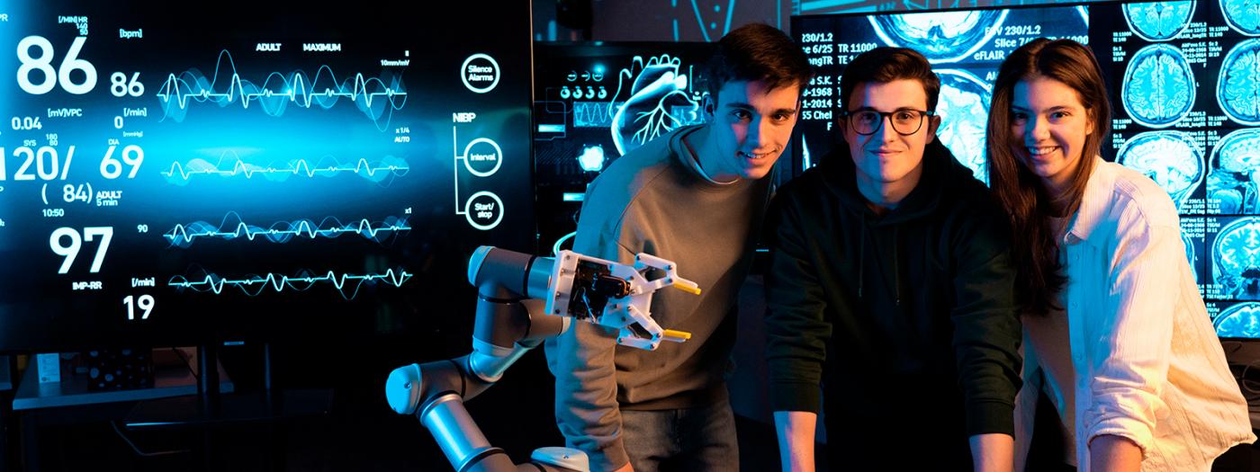
The course introduces the fundamentals of digital image processing, with a particular emphasis on its application to medical imaging. Given the central role of imaging techniques in clinical diagnosis and monitoring, the course covers the basic principles of image acquisition, representation, and processing, as well as the specific characteristics of the main medical imaging modalities.
The course provides a theoretical and practical foundation for the analysis and processing of biomedical images, including techniques for enhancement, segmentation, registration, feature extraction, and machine learning. Special emphasis is placed on the application of these methods to real-world problems, fostering students’ ability to develop computational solutions in clinical environments.
The course adopts a strongly practical approach, with sessions focused on the implementation of algorithms using Python, enabling students to consolidate their knowledge and apply it to real medical imaging data in realistic scenarios.
Titular Professors
Basic knowledge of mathematics, programming, and digital signal/image processing. Familiarity with Python is recommended.
Students acquire the knowledge and develop the skills listed below:
- Understand the concepts of digital image formation, acquisition, and basic processing in both spatial and frequency domains.
- Understand the different medical imaging modalities.
- Understand feature extraction processes for performing segmentation, detection, and classification tasks.
- Understand and apply the fundamental tools for processing medical images.
- Understand the basic functioning of machine learning and deep learning architectures applied to medical imaging.
1. Introduction to Medical Image Processing
1.1 Images
1.2 Types of images and applications
1.3 The human visual system
1.4 Digital image acquisition
1.5 Medical imaging
1.6 Image processing
2. Image Enhancement and Restoration
2.1 Image enhancement
2.2 Algebraic operations
2.3 Logical operations
2.4 Image histogram
2.5 Intensity transformation functions
2.6 Histogram transformations
2.7 Image restoration
2.8 Noise reduction
2.9 Linear filtering
2.10 Non-linear filtering
3. Image Segmentation (2D and 3D)
3.1 Image segmentation
3.2 Importance of image segmentation
3.3 Discontinuity-based segmentation
3.4 Edge detection
3.5 Thresholding
3.6 Region-based segmentation
3.7 3D image segmentation
4. Image Registration (2D and 3D)
4.1 Importance of image registration
4.2 Types of image registration
4.3 Deformable models
4.4 Rigid registration
4.5 Affine registration
4.6 Deformable registration
5. Introduction to Machine Learning
5.1 Introduction
5.2 Machine learning
5.3 Image classification
5.4 Regression
6. Introduction to Deep Learning
6.1 Introduction
6.2 Neural networks
6.3 Convolutional neural networks
6.4 Deep learning architectures in medical imaging
The teaching methodology is based on an active and theoretical-practical approach aimed at the progressive acquisition of the learning outcomes defined for the course. The subject is organized into three weekly sessions, combining lectures, theoretical capsules, practical application, and learning consolidation.
The typical structure of the sessions (except for those specifically devoted to practical work) is divided into distinct phases:
- First third: introduction of theoretical content, supported by discussion questions and practical demonstrations.
- Second third: individual or group work focused on solving practical exercises using Python or specialized medical imaging tools, based on real data and scenarios. At the end, results are shared and discussed.
- Final third: synthesis and closure of the theoretical and practical contents.
In laboratory sessions, students work individually or in groups to solve practical exercises aligned with the course content. These sessions are strategically scheduled at the end of each topic to assess concept assimilation and the ability to solve real-world problems.
The methodology integrates autonomous work, collaborative learning, and continuous formative assessment, ensuring coherence between activities, evaluation, and the workload associated with ECTS credits.
The assessment of the course is carried out through a system that evaluates both theoretical and practical content. The final grade is obtained from the following components:
- Individual theoretical-practical exam(s): 60%
- Practical laboratory sessions carried out in class: 40%
To pass the course, a minimum grade of 5 out of 10 must be obtained in both parts (theory and practice). The assessment of the practical component includes both group evaluation of the deliverables and an individual interview or exam.
If all laboratory sessions are not submitted and/or the minimum grade of 5 out of 10 is not achieved in the different assessment components, the student must take the resit examination.
The following will be assessed:
- Conceptual understanding of the theoretical foundations of medical image processing and computer vision.
- Correct application of theoretical foundations in solving theoretical-practical problems.
- Ability to implement theoretical concepts in Python.
- Rigor and coherence in the analysis of results.
- Ability to work in teams in solving real practical cases.
- Individual justification of the decisions and software solutions developed.
- R.C. Gonzalez, R.E. Woods “Digital Image Processing”, Prentice Hall, 2008
- G. Pajares, J.M. de la Cruz, “Visión por computador”, Ra-Ma
- A. de la Escalera. “Visión por computador”, Prentice Hall 2001.
- Anil K. Jain, “Fundamentals of digital image processing”, Prentice Hall, 1989
- Isaac N. Bankman, “Handbook of Medical Image Processing and Analysis”, 2009
- G. Dougherty, “Digital Image Processing for Medical Applications”, Cambridge, 2009
Optional online resources and image-processing repositories.